163 results

- Digital Images
- Online
White blood cell - polymorphonuclear leucocyte
University of Edinburgh
- Digital Images
- Online
White blood cell - polymorphonuclear leucocyte
University of Edinburgh
- Digital Images
- Online
White blood cell - polymorphonuclear leucocyte - neutrophil
University of Edinburgh
- Digital Images
- Online
TEM of leukocytes (white blood cell)
David Gregory & Debbie Marshall
- Digital Images
- Online
Human white blood cell
Anne Weston, Francis Crick Institute
- Digital Images
- Online
Human white blood cell
Anne Weston, Francis Crick Institute
- Digital Images
- Online
Blood vessel with red and white blood cells
University of Edinburgh
- Digital Images
- Online
Blood vessel with red and white blood cells
University of Edinburgh
- Archives and manuscripts
- Online
X-ray diffraction exposure referenced as "Human white blood cell"
Wilkins, Maurice Hugh Frederick, 1916-2004Date: October 1953Reference: KDBP/1/1/1194Part of: King's College London Department of Biophysics
- Books
- Online
Leucocythemia, or white cell blood : in relation to the physiology and pathology of the lymphatic glandular system. / By John Hughes Bennett.
Bennett, John Hughes, 1812-1875.Date: MDCCCLII. [1852]- Pictures
A blood droplet with a white heart monitor line against a black background representing an AIDS prevention advertisement for the AIIMS Blood Transfusion Service and NGO AIDS Cell, New Delhi. Colour lithograph, ca. 1994.
Date: ['94?]Reference: 677318i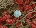
- Digital Images
- Online
Blood clot with crenated red cells
Anne Weston, Francis Crick Institute
- Digital Images
- Online
Avian blood
Royal Veterinary College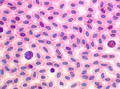
- Digital Images
- Online
Avian blood
Royal Veterinary College
- Digital Images
- Online
Blood clot
Anne Weston, Francis Crick Institute
- Digital Images
- Online
Cellular architecture of normal human skin imaged by whole mount tissue microscopy. Human skin has a rich network of white blood cells (specifically dendritic cells, T cells and macrophages) which form sheaths around blood vessels. In this image, T cells (stained for CD3; red) dendritic cells (stained for MHC class II; green) and macrophages (stained for LYVE-1; blue with some cells showing a tinge of green) can be seen. Cell nuclei have been stained with DAPI (grey). This normal cellular architecture is grossly disrupted in diseased skin (see related images). X10 magnification. Scale bar (white) represents 200 micrometres.
Dr. Xiao-nong Wang, Human Dendritic Cell Laboratory, Newcastle University
- Digital Images
- Online
Cellular architecture of normal human skin imaged by whole mount tissue microscopy. Human skin has a rich network of white blood cells (specifically dendritic cells, T cells and macrophages) which form sheaths around blood vessels. In this image, T cells (stained for CD3; red) dendritic cells (stained for MHC class II; green) and macrophages (stained for LYVE-1; blue with some cells showing a tinge of green) can be seen. Cell nuclei have been stained with DAPI (grey). This normal cellular architecture is grossly disrupted in diseased skin (see related images). X20 magnification. Scale bar (white) represents 100 micrometres.
Dr. Xiao-nong Wang, Human Dendritic Cell Laboratory, Newcastle University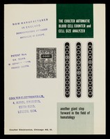
- Ephemera
- Online
The Coulter automatic blood cell counter and cell size analyzer : another giant step forward in the field of hematology / Coulter Electronics.
Coulter Electronics.Date: 1957
- Pictures
- Online
A human white blood cell (CD4) being attacked by the HIV virus; an AIDS prevention advertisement by The Ministry of Education Training, Vietnam. Colour lithograph, ca. 1995.
Date: [1995?]Reference: 678253i
- Digital Images
- Online
SEM of blood corpuscles in clot.
David Gregory & Debbie Marshall
- Digital Images
- Online
SEM of blood corpuscles in clot.
David Gregory & Debbie Marshall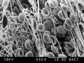
- Digital Images
- Online
Electron micrograph of blood clot,high power
David Gregory & Debbie Marshall
- Digital Images
- Online
SEM of blood corpuscles in clot.
David Gregory & Debbie Marshall
- Digital Images
- Online
SEM of blood corpuscles in clot.
David Gregory & Debbie Marshall
- Digital Images
- Online
Ultrastructure inside a macrophage cell, TEM
Kevin Mackenzie, University of Aberdeen